Implants Case Study 1648
Implants Case Study 1648
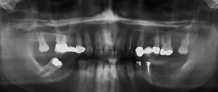
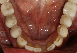
Five implants were placed in the lower arch to re-establish a functional bite.
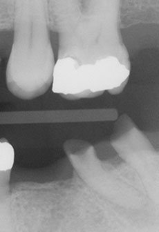
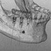
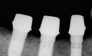
This panoramic posterior radiograph shows the two implants on the patient’s lower left (bottom right of the image) which have been completed with final restorations. The unfinished cores of the three implants on the the patient’s right can be seen on the left hand side of the image.
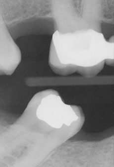
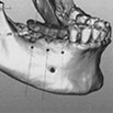
Pre Treatment Radiographs
These are the original radiographs, shown right to left. The lower molar on the right has tipped forward to the point that it wowuld be difficult to restore adequately. The decision was made to remove it. The lower left molar and the right premolar were badly decayed.
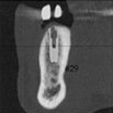
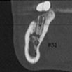
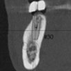
Scan Report
It’s critical that implants are placed exactly into the correct location. it is very difficult, if not impossible, to adequately restore implants that have been poorly placed. It is very important that the surgeon uses a CAT scan to determine the proper placement. Implants that have been properly placed and well restored will last indefinitely.
The image at the top of the page is the pre-treatment panoramic scan.
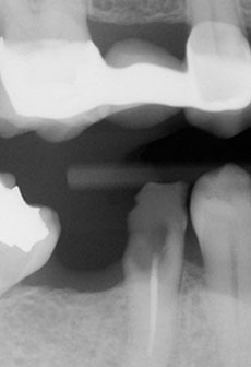
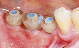
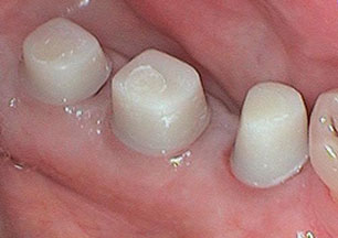
Tissue Collars and Impression Posts
Three steps in the placement of custom cores for the restoration of implants. The photograph on the left shows the healing collars which allow for expansion of the gum to create a more normal profile for the final restoration. The center photograph is the placement of the impression posts which are used by the lab to fabricate the customer cores shown in the third image. It’s important to determine that the cores have been adequately seated on the implants.
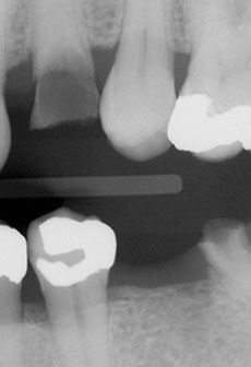
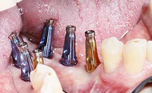
Core Placement
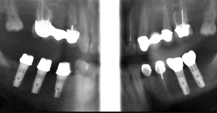
The cores are held in place by screws that attach to the implant through the core. The hole for the screw is then filled with composite. It’s possible to take out and replace the core at some point in the future by removing the composite and unscrewing the screw. The center radiograph shows the final cementation of the restorations on top of the cores.
This panoramic posterior radiograph shows the two implants on the patient’s lower left (bottom right of the image) which have been completed with final restorations. The unfinished cores of the three implants on the the patient’s right can be seen on the left hand side of the image.
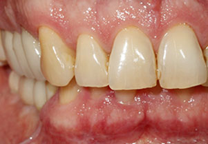
Post Treatment Photos
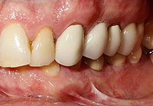
A total of five implants were placed to restore missing and decayed lower posterior teeth. These final photographs were taken following cementation fo the lower right implants, shown in the right hand photograph. The lower left implants were placed earlier.
Case Studies
- Vertical Dimension Case Study 99463
- Reconstruction Case Study 99461
- Periodontal Disease Case Study 99469
- Partial Reconstruction Case Study 99466
- Oral Rehabilitation Case Study 2540
- Maxillary Case Study 99459
- Implants Case Study 3116
- Implants Case Study 1648
- Implants Case Study 1443
- Implants Case Study 1169
- Full Restoration Case Study 99465
- Full Mouth Cosmetics
- Front Teeth Restoration
- Fluorsis
- Cosmetic Tissue Control
- Oh Canada
- Airway Obstruction
- Temporomandibular Joint Dysfunction (TMJ)